What Does The Brain Do? (Lobes And Regions)

An illustrated handout designed for psychoeducation explaining the functions of each lobe of the human brain. Limbic regions such such as the cingular, hippocampus, and amygdala are also included.

Download or send

Professional version

Offers theory, guidance, and prompts for mental health professionals. Downloads are in Fillable PDF format where appropriate.

Client version

Includes client-friendly guidance. Downloads are in Fillable PDF format where appropriate.

Editable version (PPT)

An editable Microsoft PowerPoint version of the resource.
Overview

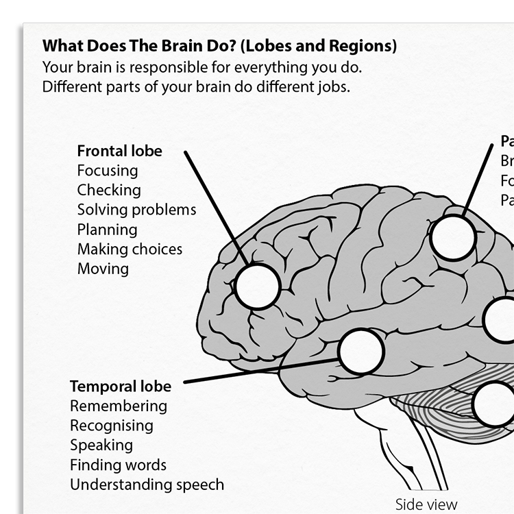
The brain is the central organ of the body’s nervous system and is essential for processes such as perception, movement, language, and memory. This illustrated handout provides a concise summary of the cerebral cortex, outlining the distinct functions of its four lobes - frontal, parietal, temporal, and occipital. It also introduces other important brain structures, including the limbic system, which plays a key role in emotional processing and memory.

Why Use This Resource?

This information handout assists therapists in explaining brain anatomy and function to clients. Understanding brain function can normalize symptoms and enhance therapeutic engagement.

  • Informs clients about the brain’s structure and its role in cognition and emotion.
  • Helps therapists in linking symptoms with brain function.
  • Supports conversations on brain injuries and related cognitive impairments.

Key Benefits

Understanding

Helps clients grasp how different brain regions contribute to behavior and cognition.

Education

Provides clear, accessible information for clients with neurological impairments.

Engagement

Motivates clients by linking symptoms to brain structure.

Support

Assists therapists in demonstrating effects of brain injuries on mental health.

Who is this for?

Memory Problems

Helps clients understand how different brain regions contribute to memory formation, storage, and recall. Useful in contexts such as mild cognitive impairment, dementia, brain injury, and trauma-related memory disruptions.

Traumatic Brain Injury (TBI)

Explain cognitive changes post-injury, focusing on areas like executive function and emotional regulation.

Stroke

Address impairments in movement, speech, or other affected domains.

Neurological Disorders

Educate about brain damage and cognitive symptoms post-infection or accident.

Anxiety

Emotional responses and their relation to limbic system activity.

Integrating it into your practice

01

Discuss

Open conversations about the client's specific brain injury or mental health condition.

02

Illustrate

Use visual aids from the handout to demonstrate affected brain areas.

03

Explain

Clarify how brain activity or damage translates to cognitive and emotional symptoms.

04

Reflect

Facilitate client reflection on how brain function affects their experience.

Theoretical Background & Therapist Guidance

Understanding how different regions of the brain contribute to cognitive, emotional, and behavioural processes is important in many therapeutic approaches. Cognitive neuroscience has provided insights into how specific brain structures support thinking, emotion, and behaviour. Over the past forty years, advances in neuroimaging have made it possible to observe how patterns of brain activity correlate with psychological phenomena (Vincent et al., 2008; Ham et al., 2014). These developments have enabled clinicians and researchers to better understand the links between brain function and the symptoms experienced by individuals, whether those symptoms arise from neurological injury or mental health conditions.

This handout introduces clients to the major anatomical regions of the brain, particularly the four lobes of the cerebral cortex - frontal, parietal, temporal, and occipital - as well as the limbic system. Each region plays a distinct role. The frontal lobes are central to high-level executive processes such as planning, reasoning, decision-making, and emotion regulation (Mateer & Sira, 2006; Miyake et al., 2000).

The temporal lobes are essential for memory, auditory processing, and language comprehension (Ogar et al., 2011; Scott, 2005). The parietal lobes help integrate sensory information and are critical for spatial awareness, attention, and mathematical reasoning (Ham et al., 2014). The occipital lobes are primarily involved in visual processing and recognition (Biran & Coslett, 2003). The limbic system - which includes the amygdala, hippocampus, and cingulate cortex - plays a central role in emotional processing, memory formation, and the regulation of emotional responses (Gupta et al., 2011; Leech & Sharp, 2014).

Damage to specific brain regions - whether from stroke, traumatic brain injury, infection, or degenerative illness - can lead to characteristic cognitive and emotional difficulties. Individuals with damage to the frontal lobes may have trouble initiating or organising tasks and regulating emotional responses (Mateer & Sira, 2006). Temporal lobe involvement can manifest as memory problems, language disturbances, or difficulty recognising familiar voices and faces (Bird & Burgess, 2008; Dellacherie et al., 2011). Disruption to the limbic system is particularly relevant to emotional dysregulation. In post-traumatic stress disorder, for example, increased activity in the amygdala and reduced regulation from frontal regions have been associated with heightened emotional reactivity, fear responses, and persistent intrusive memories (Ham et al., 2014; Gupta et al., 2011).

Psychoeducation about the structure and function of the brain can play an important role in therapy. Helping clients understand their symptoms in terms of brain processes can normalise distressing experiences, reduce self-blame, and support therapeutic engagement. This handout is designed to facilitate such conversations, offering a simple yet scientifically grounded overview of brain function that can be adapted for use with a wide range of clients.

What's inside

  • Illustrated descriptions of the brain's lobes and regions.
  • Visual aids for client education.
  • Guidance on using the handout for psychoeducation.
Get access to this resource

FAQs

Use it to explain brain function and structure, especially for clients with conditions tied to brain changes, enhancing understanding and engagement.
Yes, its simplified design and language make it accessible for various clients, including those with neurological impairments.
Absolutely. Incorporate it during assessments and therapy sessions to discuss the neurobiological aspects of individual symptoms.

How This Resource Improves Clinical Outcomes

Enhancing client understanding of their neurological and psychological symptoms through education encourages active participation in therapy:

  • Clients understand their symptoms in relation to brain function.
  • Clients can be motivated by insights into their condition.
  • Facilitates clearer discussions between clinician and client.

Therapists benefit by having a structured tool for explaining complex concepts, reinforcing the therapeutic process through informed discussion.

References And Further Reading

  • Biran, I., & Coslett, H. B. (2003). Visual agnosia. Current Neurology and Neuroscience Reports, 3(6), 508-512.
  • Bird, C. M., & Burgess, N. (2008). The hippocampus and memory: insights from spatial processing. Nature Reviews Neuroscience, 9(3), 182-194.
  • Carrera, E., & Bogousslavsky, J. (2006). The thalamus and behavior: effects of anatomically distinct strokes. Neurology, 66(12), 1817-1823.
  • Dellacherie, D., Hasboun, D., Baulac, M., Belin, P., & Samson, S. (2011). Impaired recognition of fear in voices and reduced anxiety after unilateral temporal lobe resection. Neuropsychologia, 49(4), 618-629.
  • Gupta, R., Koscik, T. R., Bechara, A., & Tranel, D. (2011). The amygdala and decision-making. Neuropsychologia, 49(4), 760-766.
  • Ham, T. E., Bonnelle, V., Hellyer, P., Jilka, S., Robertson, I. H., Leech, R., & Sharp, D. J. (2014). The neural basis of impaired self-awareness after traumatic brain injury. Brain, 137(2), 586-597.
  • Hirshorn, E. A., & Thompson-Schill, S. L. (2006). Role of the left inferior frontal gyrus in covert word retrieval: neural correlates of switching during verbal fluency. Neuropsychologia, 44(12), 2547-2557.
  • Karnath, H. O., Rüter, J., Mandler, A., & Himmelbach, M. (2009). The anatomy of object recognition-visual form agnosia caused by medial occipitotemporal stroke. Journal of Neuroscience, 29(18), 5854-5862.
  • Leech, R., & Sharp, D. J. (2014). The role of the posterior cingulate cortex in cognition and disease. Brain, 137(1), 12-32.
  • Miyake, A., Friedman, N. P., Emerson, M. J., Witzki, A. H., Howerter, A., & Wager, T. D. (2000). The unity and diversity of executive functions and their contributions to complex “frontal lobe” tasks: A latent variable analysis. Cognitive Psychology, 41(1), 49-100.
  • Manto, M.U. (2010) Symptoms of Cerebellar Disorders, Ch.3 in: Cerebellar Disorders: A Practical Approach to Diagnosis and Management. Cambridge University Press, UK.
  • Mateer, C. A., & Sira, C. S. (2006). Cognitive and emotional consequences of TBI: intervention strategies for vocational rehabilitation. NeuroRehabilitation, 21(4), 315-326.
  • Ogar, J. M., Baldo, J. V., Wilson, S. M., Brambati, S. M., Miller, B. L., Dronkers, N. F., & Gorno-Tempini, M. L. (2011). Semantic dementia and persisting Wernicke’s aphasia: linguistic and anatomical profiles. Brain and Language, 117(1), 28-33.
  • Scott, S. K. (2005). Auditory processing—speech, space and auditory objects. Current Opinion In Neurobiology, 15(2), 197-201.
  • Vincent, J. L., Kahn, I., Snyder, A. Z., Raichle, M. E., & Buckner, R. L. (2008). Evidence for a frontoparietal control system revealed by intrinsic functional connectivity. Journal of Neurophysiology, 100(6), 3328-3342.